
VGG19 in MRI Brain Tumor Classification: A Systematic Review of Validation Rigor and Clinical Readiness
Keywords:
Brain tumor classification, magnetic resonance imaging, VGG19, transfer learning, systematic review, explainable artificial intelligenceAbstract
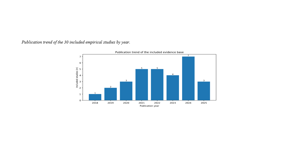
VGG19 remains a widely used reference backbone for MRI-based brain tumor classification; however, the literature is difficult to interpret because model modifications, dataset provenance, data-splitting strategies, and evaluation practices vary substantially. This systematic review provides a VGG19-centered evidence map linking architectural adaptations to validation rigor and indicators of clinical readiness. The review was updated in accordance with PRISMA 2020 principles. Searches were conducted in PubMed, IEEE Xplore, ScienceDirect, and SpringerLink, supplemented by citation searching, to identify peer-reviewed studies published from January 2015 through December 2025; the search was last updated on July 27, 2026. Because the original database export files were not retained, database-specific retrieval and duplicate counts are not reported. The final evidence base was reconciled at the reference level: 50 unique references were classified, 20 contextual or methodological sources were separated, and 30 empirical studies were included. Data were extracted on classification task, MRI source, class structure, role of VGG19, preprocessing procedures, splitting unit, validation design, performance metrics, interpretability, and reproducibility. Reporting quality was appraised using the CLAIM 2024 domains. Results were not statistically pooled because of substantial outcome heterogeneity; instead, a quantitative descriptive synthesis and structured comparative evidence map were used. Research output increased after 2021. VGG19 was employed as a stand-alone classifier, transfer-learning feature extractor, fine-tuned backbone, hybrid-model component, ensemble member, and component of explainable and federated-learning frameworks. All 30 studies reported accuracy or an accuracy-derived outcome; 25 reported class-specific metrics, 12 employed cross-validation, six included external or distributed validation, seven incorporated explainability methods, and four provided code or other implementation materials. High classification accuracies were frequently reported for Figshare and related contrast-enhanced T1-weighted MRI datasets; however, the strength of the evidence was often limited by image-level data splitting, incomplete reporting of patient provenance, lack of calibration assessment, and limited multicenter validation. The principal contribution of this review is a three-axis synthesis demonstrating that architectural improvements should be interpreted jointly with validation rigor and evidence of clinical readiness. Fine-tuned and hybrid VGG19-based systems represent competitive baselines; however, reliable clinical translation requires patient-level data separation, external validation, class-specific performance reporting, calibration assessment, explainability, and transparent documentation of computational methods and implementation.
Downloads
References
1. Menze BH, Jakab A, Bauer S, Kalpathy-Cramer J, Farahani K, Kirby J, et al. The Multimodal Brain Tumor Image Segmentation Benchmark (Brats). IEEE Transactions on Medical Imaging. 2015;34(10):1993-2024. [PMID: 25494501] [DOI]
2. Bakas S, Akbari H, Sotiras A, Bilello M, Rozycki M, Kirby JS, et al. Advancing The Cancer Genome Atlas Glioma MRI Collections With Expert Segmentation Labels And Radiomic Features. Scientific Data. 2017;4:170117. [PMID: 28872634] [DOI]
3. Litjens G, Kooi T, Bejnordi BE, Setio AAA, Ciompi F, Ghafoorian M, et al. A Survey On Deep Learning In Medical Image Analysis. Medical Image Analysis. 2017;42:60-88. [PMID: 28778026] [DOI]
4. Zhou SK, Greenspan H, Davatzikos C, Duncan JS, Van Ginneken B, Madabhushi A, et al. A Review Of Deep Learning In Medical Imaging: Imaging Traits, Technology Trends, Case Studies With Progress Highlights, And Future Promises. Proceedings of the IEEE. 2021;109(5):820-38. [PMID: 37786449] [PMCID: PMC10544772] [DOI]
5. Cheng J, Huang W, Cao S, Yang R, Yang W, Yun Z, et al. Enhanced Performance Of Brain Tumor Classification Via Tumor Region Augmentation And Partition. PLoS ONE. 2015;10(10):e0140381. [PMID: 26447861] [DOI]
6. Khan HA, Jue W, Mushtaq M, Mushtaq MU. Brain Tumor Classification In MRI Image Using Convolutional Neural Network. Mathematical Biosciences and Engineering. 2020;17(5):6203-16. [PMID: 33120595] [DOI]
7. Alanazi MF, Ali MU, Hussain SJ, Zafar A, Mohatram M, Irfan M, et al. Brain Tumor/Mass Classification Framework Using Magnetic-Resonance-Imaging-Based Deep Learning. Sensors. 2022;22(1):372. [PMID: 35009911] [PMCID: PMC8749789] [DOI]
8. Noreen N, Palaniappan S, Qayyum A, Ahmad I, Alassafi MO. Brain Tumor Classification Based On Fine-Tuned Models And The Ensemble Method. Computers, Materials & Continua. 2021;67(3):3967-82. [DOI]
9. Islam MM, Barua P, Rahman M, Ahammed T, Akter L, Uddin J. Transfer Learning Architectures With Fine-Tuning For Brain Tumor Classification Using Magnetic Resonance Imaging. Healthcare Analytics. 2023;4:100270. [DOI]
10. Rasa SM, Islam MM, Talukder MA, Uddin MA, Khalid M, Kazi M, et al. Brain Tumor Classification Using Fine-Tuned Transfer Learning Models On Magnetic Resonance Imaging (MRI) Images. Digital Health. 2024;10. [PMID: 39381813] [PMCID: PMC11459499] [DOI]
11. Rastogi D, Johri P, Donelli M, Kumar L, Bindewari S, Raghav A, et al. Brain Tumor Detection And Prediction In MRI Images Utilizing A Fine-Tuned Transfer Learning Model Integrated Within Deep Learning Frameworks. Life. 2025;15(3):327. [PMID: 40141673] [PMCID: PMC11944010] [DOI]
12. Khan AR, Khan S, Harouni M, Abbasi R, Iqbal S, Mehmood Z. Brain Tumor Segmentation Using K-Means Clustering And Deep Learning With Synthetic Data Augmentation For Classification. Microscopy Research and Technique. 2021;84(7):1389-99. [PMID: 33524220] [DOI]
13. Rasool M, Ismail NA, Boulila W, Ammar A, Samma H, Yafooz WMS, et al. A Hybrid Deep Learning Model For Brain Tumour Classification. Entropy. 2022;24(6):799. [PMID: 35741521] [PMCID: PMC9222774] [DOI]
14. Veeramuthu A, Meenakshi S, Mathivanan G, Kotecha K, Saini JR, Vijayakumar V, et al. MRI Brain Tumor Image Classification Using A Combined Feature And Image-Based Classifier. Frontiers in Psychology. 2022;13:848784. [PMID: 35310201] [PMCID: PMC8931531] [DOI]
15. Aurna NF, Yousuf MA, Taher KA, Azad AKM, Moni MA. A Classification Of MRI Brain Tumor Based On Two Stage Feature Level Ensemble Of Deep Cnn Models. Computers in Biology and Medicine. 2022;146:105539. [PMID: 35483227] [DOI]
16. Nassar SE, Yasser I, Amer HM, Mohamed MA. A Robust MRI-Based Brain Tumor Classification Via A Hybrid Deep Learning Technique. The Journal of Supercomputing. 2024;80:2403-27. [DOI]
17. Tummala S, Kadry S, Bukhari SAC, Rauf HT. Classification Of Brain Tumor From Magnetic Resonance Imaging Using Vision Transformers Ensembling. Current Oncology. 2022;29(10):7498-511. [PMID: 36290867] [DOI]
18. Hossain S, Chakrabarty A, Gadekallu TR, Alazab M, Piran MJ. Vision Transformers, Ensemble Model, And Transfer Learning Leveraging Explainable Ai For Brain Tumor Detection And Classification. IEEE Journal of Biomedical and Health Informatics. 2024;28(3):1261-72. [PMID: 37043319] [DOI]
19. Rohini A, Praveen C, Mathivanan SK, Muthukumaran V, Mallik S, Alqahtani MS, et al. Multimodal Hybrid Convolutional Neural Network Based Brain Tumor Grade Classification. BMC Bioinformatics. 2023;24:382. [PMID: 37817066] [PMCID: PMC10566188] [DOI]
20. Ullah MS, Khan MA, Albarakati HM, Damaševičius R, Alsenan S. Multimodal Brain Tumor Segmentation And Classification From MRI Scans Based On Optimized Deeplabv3+ And Interpreted Networks Information Fusion Empowered With Explainable Ai. Computers in Biology and Medicine. 2024;182:109183. [PMID: 39357134] [DOI]
21. Selvaraju RR, Cogswell M, Das A, Vedantam R, Parikh D, Batra D. Grad-Cam: Visual Explanations From Deep Networks Via Gradient-Based Localization. Proceedings of the IEEE International Conference on Computer Vision (ICCV)2017. p. 618-26. [DOI]
22. Woo S, Park J, Lee JY, Kweon IS. Cbam: Convolutional Block Attention Module. Proceedings of the European Conference on Computer Vision (ECCV)2018. p. 3-19. [DOI]
23. Haque R, Hassan MM, Bairagi AK, Islam SMS. Neuronet19: An Explainable Deep Neural Network Model For The Classification Of Brain Tumors Using Magnetic Resonance Imaging Data. Scientific Reports. 2024;14:1524. [PMID: 38233516] [PMCID: PMC10794704] [DOI]
24. Alshuhail A, Thakur A, Chandramma R, Mahesh TR, Almusharraf A, Vinoth Kumar V, et al. Refining Neural Network Algorithms For Accurate Brain Tumor Classification In MRI Imagery. BMC Medical Imaging. 2024;24:118. [PMID: 38773391] [PMCID: PMC11110259] [DOI]
25. Kothadiya D, Rehman A, Alghofaily B, Bhatt C, Ayesha N, Saba T. Vgx: Vgg19-Based Gradient Explainer Interpretable Architecture For Brain Tumor Detection In Microscopy Magnetic Resonance Imaging (Mmri). Microscopy Research and Technique. 2025;88(5):1544-54. [PMID: 39825619] [DOI]
26. Nawaz M, Nazir T, Masood M, Mehmood A, Mahum R, Khan MA, et al. Analysis Of Brain MRI Images Using Improved Cornernet Approach. Diagnostics. 2021;11(10):1856. [PMID: 34679554] [PMCID: PMC8535141] [DOI]
27. Collins GS, Reitsma JB, Altman DG, Moons KGM. Transparent Reporting Of A Multivariable Prediction Model For Individual Prognosis OR Diagnosis: Tripod. Annals of Internal Medicine. 2015;162(1):55-63. [PMID: 25560714] [DOI]
28. Page MJ, Mckenzie JE, Bossuyt PM, Boutron I, Hoffmann TC, Mulrow CD, et al. The Prisma 2020 Statement: An Updated Guideline For Reporting Systematic Reviews. BMJ. 2021;372:n71. [PMID: 33782057] [DOI]
29. Tejani AS, Klontzas ME, Gatti AA, Mongan JT, Moy L, Park SH, et al. Checklist For Artificial Intelligence In Medical Imaging (Claim): 2024 Update. Radiology: Artificial Intelligence. 2024;6(4):e240300. [DOI]
30. Lekadir K, Frangi AF, Porras AR, Glocker B, Cintas C, Langlotz CP, et al. Future-Ai: International Consensus Guideline For Trustworthy And Deployable Artificial Intelligence In Healthcare. BMJ. 2025;388:e081554. [DOI]
Downloads
Publication Timeline
- Submitted
- Revised
- Accepted