Effects of Pilates Training and Vitamin D Supplementation on BMD, BDNF, and Mineral Status in Women with Osteosarcoma
Abstract
Objective: Osteosarcoma and its treatment protocols may disrupt bone mineral homeostasis, reduce bone mineral density (BMD), and alter neurotrophic factors such as brain-derived neurotrophic factor (BDNF). This study aimed to investigate the effects of an 8-week Pilates training program combined with vitamin D supplementation on serum mineral-related biomarkers, BDNF, and BMD in women with osteosarcoma.
Methods: Fifty women with medically stable osteosarcoma (age: 29.50 ± 0.79 years; BMI: 23.65 kg/m²) participated in this randomized controlled trial. After screening and eligibility assessment, participants were randomly assigned to five groups (n = 10/group): Pilates + vitamin D (ExD), Pilates + placebo (ExP), Pilates only (Ex), vitamin D only (D), and control (CO). The Pilates intervention consisted of supervised sessions three times per week for 8 weeks (24 sessions total). Participants in supplementation groups received 5000 IU vitamin D3 weekly for 8 weeks. Supplement/placebo administration and outcome assessment were blinded.
Results: Eight weeks of combined Pilates and vitamin D supplementation were associated with significant improvements in serum calcium (p = 0.003), magnesium (p = 0.002), phosphorus (p = 0.01), BDNF (p = 0.01), and BMD (p = 0.01) compared with control conditions. Post-hoc analyses indicated that the ExD group demonstrated greater improvements than control and single-intervention groups (p ≤ 0.05).
Conclusion: Combined Pilates training and vitamin D supplementation may improve serum mineral-related biomarkers, BDNF, and BMD-related outcomes in women with osteosarcoma. Larger and longer-term studies are needed to confirm these preliminary findings.
Introduction
Modern rehabilitation strategies increasingly emphasize the integration of targeted exercise programs with nutritional interventions to optimize bone health and recovery outcomes. Among these, the combination of structured physical activity including low-impact modalities such as Pilates and vitamin D supplementation has gained attention for its potential complementary effects on bone metabolism, mineral homeostasis, and neurotrophic factors such as brain-derived neurotrophic factor (BDNF). This integrative approach may be particularly relevant in clinical populations with compromised skeletal integrity, including patients with osteosarcoma.
Osteosarcoma is the most common primary malignant bone tumor, predominantly affecting adolescents and young adults. It is characterized by aggressive bone destruction, altered bone remodeling, and significant impairments in bone mineral density (BMD). In addition to the direct effects of the tumor, treatments such as chemotherapy and surgical resection further exacerbate bone loss, reduce physical function, and impair quality of life. Consequently, identifying safe and effective rehabilitation strategies that can improve bone metabolism and functional outcomes in these patients is of considerable clinical importance (1).
Vitamin D plays a central role in regulating calcium–phosphorus homeostasis and bone remodeling. It enhances intestinal calcium absorption and contributes to osteoblast and osteoclast regulation (2). In patients with cancer, including osteosarcoma, vitamin D deficiency is relatively common and has been associated with poorer bone health and increased fracture risk (3). However, systematic reviews indicate that its effect on BMD is inconsistent across populations, particularly when used as a standalone intervention (4, 5). These findings highlight the importance of combining supplementation with other interventions, such as exercise.
Exercise, particularly weight-bearing and resistance-based activity, is widely recognized as a key stimulus for bone adaptation. Mechanical loading can stimulate osteogenic responses and contribute to improvements in bone strength and musculoskeletal function (6, 7). Structured exercise interventions in cancer populations have been associated with improvements in physical function, musculoskeletal health, and potentially bone-related outcomes, although evidence remains heterogeneous (8). Pilates, as a low-impact and controlled exercise modality, may represent a feasible option in clinical populations due to its emphasis on postural control, flexibility, and neuromuscular coordination.
Recent evidence suggests that structured exercise approaches, including low-impact modalities, may improve balance, muscle strength, and functional mobility, which are critical for reducing fall risk and maintaining skeletal integrity (8). In addition to skeletal adaptations, exercise has also been associated with changes in neurotrophic factors such as BDNF, which plays an important role in neuronal function and may also be involved in bone-related processes (9, 10).
Emerging evidence suggests that vitamin D may interact with neurobiological pathways, including those related to BDNF regulation, although these mechanisms are not fully understood (11). This raises the possibility of complementary effects between exercise and vitamin D in influencing both skeletal and neurophysiological outcomes. However, despite these promising findings, there is limited research specifically examining the combined effects of Pilates training and vitamin D supplementation in osteosarcoma patients.
Another important consideration is the role of inflammation in bone metabolism among cancer patients. Chronic inflammation may disrupt bone remodeling by increasing osteoclast activity and inhibiting osteoblast function (12).
In summary, the integration of structured exercise and vitamin D supplementation represents a potentially valuable approach for improving bone health and neurobiological function in osteosarcoma patients. Therefore, the present study aims to investigate the effects of an 8-week Pilates training program combined with vitamin D supplementation on mineral-related biomarkers, BDNF levels, and bone mineral density in women with osteosarcoma.
Materials and Methods
The present study was approved by the Ethics Committee of Shahid Chamran University of Ahvaz (IR.SCU.REC.1404.147). The research was conducted in accordance with ethical principles for medical research, and all participants provided written informed consent before participation.
Participants
After the invitation to participate in the study, 100 women were screened. After review of the inclusion criteria, 80 individuals were eligible. Finally, 50 participants were randomly selected and allocated to the five study groups. Participants with osteosarcoma (body mass index: 23.65 kg/m², age: 29.50 ± 0.79 years, and weight: 62.14 ± 1.31 kg) were recruited through a call announcement at Sadr Clinic in Tehran.
The inclusion criteria were as follows: non-smoking status, no regular physical activity during the previous year (>2 hours/week), ability to perform physical exercise, at least four years since osteosarcoma diagnosis, and at least two years since bone marrow transplantation, where applicable. Participants were excluded if they met any of the following criteria: (1) failure to follow the researchers’ recommendations, (2) irregular attendance (less than 80% of scheduled exercise sessions), (3) changes in the type or dosage of prescribed medications during the intervention period, or (4) withdrawal of consent or unwillingness to continue the intervention.
After complete explanations about the research process and the potential advantages and disadvantages of the study, the Physical Activity Readiness Questionnaire (PAR-Q) was completed to assess medical history and participants’ condition during the intervention. Sample size was calculated using G*Power software based on a randomized pre-test–post-test design with five groups, a confidence level of 95%, statistical power of 80%, and a medium effect size (based on available prior exercise-intervention effect-size estimates due to limited osteosarcoma-specific data), resulting in 50 eligible participants. Participants were then randomly divided into five groups: exercise + vitamin D (ExD, n = 10), exercise + placebo (ExP, n = 10), exercise only (Ex, n = 10), vitamin D only (D, n = 10), and control (CO, n = 10). Supplement/placebo administration and outcome assessment were blinded. The placebo was prepared to be similar to the vitamin D supplement to reduce the effect of participant awareness of the intervention.
During the study, the dropout rate of participants was carefully monitored. Any participant who did not attend at least 80% of the sessions or did not take the supplement/placebo regularly was excluded from the final analysis. The average adherence across groups was over 90%, and no serious adverse events were reported.
Study Procedure
This study used an 8-week intervention period consisting of 24 Pilates sessions (3 sessions/week). Pre-test assessments were conducted before randomization, and post-test assessments were performed after completion of the 8-week intervention under comparable conditions.
Exercise training
The Pilates exercise program was designed for 8 weeks, three sessions per week (24 sessions), based on a prior Pilates-related intervention study (4). Session duration increased from 40 minutes in Weeks 1–2 to 55 minutes in Weeks 7–8, including warm-up and cool-down (4). Exercise intensity was measured using the rating of perceived exertion (RPE) based on the traditional Borg 6–20 scale (4). In this scale, 6 corresponds to “no exertion at all” and 20 corresponds to “maximal exertion”; RPE 9–10 was used for warm-up, RPE 14–16 for the main exercises, and RPE 9–10 for cool-down. Participants were instructed to stop each exercise before the final 3–4 repetitions at the difficult level equivalent to RPE 16. To determine and control exercise intensity, a percentage of maximum heart rate (HRmax) was used. Before the 8-week intervention, all participants underwent an HRmax assessment test. The test was conducted on a cycle ergometer (Monark 828E, Sweden) under the supervision of a physician and an exercise physiologist. After a 5-minute warm-up at 25 watts with a pedaling rate of 50–60 rpm, workload was increased by 25 watts every 2 minutes until volitional exhaustion or until the participant could no longer maintain a pedaling rate above 50 rpm. Heart rate was continuously recorded using a Polar H10 chest strap (Polar Electro, Finland), and the highest heart rate achieved during the test was recorded as HRmax. Only exercise-assigned groups performed Pilates. Exercise intensity was gradually increased according to the principle of overload: Weeks 1–3 were performed at 50–55% HRmax, Weeks 4–6 at 55–60% HRmax, and Weeks 7–8 at 60–65% HRmax. Real-time data recorded by smartwatches during exercise allowed continuous monitoring and adjustment of activity intensity according to each participant’s characteristics (see Supplementary Table 1).
Training Variables | Week 1 | Week 2 | Week 3 | Week 4 | Week 5 | Week 6 | Week 7 | Week 8 |
|---|---|---|---|---|---|---|---|---|
Number of sessions/week | 3 | 3 | 3 | 3 | 3 | 3 | 3 | 3 |
Number of sets | 3 | 3 | 3 | 3 | 3 | 3 | 3 | 3 |
Repetitions | 4 | 4 | 5 | 5 | 6 | 6 | 10 | 10 |
Number of exercises | 10 | 10 | 11 | 11 | 12 | 12 | 13 | 13 |
RPE | 14 | 14 | 14 | 15 | 15 | 15 | 15 | 16 |
Intensity (% HRmax) | 50–55 | 50–55 | 50–55 | 55–60 | 55–60 | 55–60 | 60–65 | 60–65 |
Rest time (s) | 30–60 | 30–60 | 30–60 | 30–60 | 30–60 | 30–60 | 30–60 | 30–60 |
Rest between sets (min) | 4 | 4 | 4 | 4 | 4 | 3 | 3 | 3 |
Total session duration (min) | 40 | 40 | 45 | 45 | 50 | 50 | 55 | 55 |
Warm-up (min) | 10 | 10 | 10 | 10 | 10 | 10 | 10 | 10 |
Cool-down (min) | 10 | 10 | 10 | 10 | 10 | 10 | 10 | 10 |
Vitamin D
Patients in supplementation groups received 5000 IU vitamin D3 orally once weekly for 8 weeks under physician supervision. For consistency, vitamin D status was classified throughout the manuscript as:
- Deficiency: <10 ng/mL
- Insufficiency: 10–30 ng/mL
- Sufficiency: ≥30 ng/mL (14).
Body Composition and Anthropometric Measures
Demographic parameters (age, alcohol consumption, smoking status, and family history of disease) and body-composition measures (height, weight, and BMI) were recorded before and after the intervention. Age, alcohol consumption, smoking status, family history of disease, and other health characteristics were obtained using a questionnaire. Body composition and physiological variables were measured according to the guidelines of the International Biological Program (13). BMI was calculated by dividing weight (kg) by height squared (m²) (see Supplementary Table 2 for descriptive data).
Characteristics of participants | Groups | Pre-Test | Post-Test | P-value | ||
|---|---|---|---|---|---|---|
Mean | SD | Mean | SD | |||
Age (years) | Control | 29.70 | .15 | - | - | 0.2 |
Vitamin D | 29.10 | .40 | - | - | ||
Exercise | 29.70 | .21 | - | - | ||
Exercise + placebo | 29.40 | .22 | - | - | ||
exercise-Vitamin D | 29.60 | .16 | - | - | ||
Height (cm) | Control | 162.5 | 6.2 | - | - | 0.9 |
Vitamin D | 163.33 | 3.2 | - | - | ||
Exercise | 163.5 | 2.3 | - | - | ||
Exercise + placebo | 162.2 | 1.5 | - | - | ||
exercise-Vitamin D | 164.7 | 2.31 | - | - | ||
Weight (kg) | Control | 61.80 | .46 | 62.50 | .45 | 0.5 |
Vitamin D | 62.70 | .53 | 62.40 | .42 | ||
Exercise | 62.00 | .33 | 62.50 | .50 | ||
Exercise + placebo | 62.00 | .44 | 61.60 | .37 | ||
exercise-Vitamin D | 62.20 | .24 | 62.90 | .40 | ||
BMI (kg.m2) | Control | 23.4 | 0.46 | 23.66 | 0.45 | 0.5 |
Vitamin D | 23.5 | 0.53 | 23.4 | 0.42 | ||
Exercise | 23.57 | 0.33 | 23.75 | 0.50 | ||
Exercise + placebo | 23.57 | 0.44 | 23.41 | 0.37 | ||
exercise-Vitamin D | 22.95 | 0.24 | 23.20 | 0.40 | ||
* No statistically significant between-group differences were observed for baseline characteristics (P > 0.05). Data normality was assessed using the Kolmogorov–Smirnov test. | ||||||
Bone Mineral Density Assessment
Bone mineral density (BMD) was assessed using dual-energy X-ray absorptiometry (DXA) (Hologic Inc., Marlborough, MA, USA). Lumbar spine BMD (L1–L4) was measured by a trained and certified technician following the manufacturer's standardized scanning protocol. All participants underwent BMD assessment at baseline before randomization and again after completion of the 8-week intervention under identical measurement conditions. Participants were scanned wearing light clothing without metal objects, and the same DXA scanner was used for all measurements. The DXA system was calibrated daily according to the manufacturer's quality-control recommendations, and all scans were performed by the same operator to minimize inter-operator variability. BMD values were expressed in grams per square centimeter (g/cm²).
Blood Sampling and Analysis
Blood samples were collected at two time points: 48 hours before the intervention and 48 hours after completion of the final intervention session. At each time point, 5 mL of blood was collected from the brachial vein after an overnight fast under physician supervision. Samples were centrifuged at 3500 rpm, and serum was separated and stored at −20°C until analysis. All blood samples were obtained between 8:00 and 9:00 a.m. to minimize circadian and nutritional variability.
Samples were allowed to clot at room temperature and were centrifuged at 3500 rpm for 10 minutes. Serum was separated and stored at −20°C until biochemical analysis. To minimize analytical variability, all samples from each participant were analyzed within the same assay batch, and measurements were performed in duplicate whenever possible.
Serum calcium, magnesium, and phosphorus concentrations were measured using commercially available colorimetric assay kits (Elabscience Biotechnology Co., Wuhan, China) according to the manufacturer’s instructions. Serum BDNF concentrations were determined using a commercially available enzyme-linked immunosorbent assay (ELISA) kit (Elabscience Biotechnology Co., Wuhan, China). The intra- and inter-assay coefficients of variation (CVs) reported by the manufacturer were <10% and <12%, respectively.
Because serum BDNF concentrations may be influenced by platelet degranulation during the clotting process, BDNF findings should be interpreted cautiously. No repeated freeze–thaw cycles were performed prior to biochemical analysis.
Statistical Method
Statistical analyses were performed using SPSS software, version 26 (IBM Corp., Armonk, NY, USA). Descriptive statistics were applied to determine the mean and standard deviation (SD) of all measured variables. The Kolmogorov–Smirnov test was used to evaluate the normality of data distribution. To compare the effects between groups, we performed Bonferroni post-hoc tests following the two-way mixed ANOVA. Specifically, we made two sets of comparisons: (1) each intervention group (D, Ex, ExP, and ExD) versus the Control group to assess the effect of each intervention relative to no treatment, and (2) the ExD group versus each of the other intervention groups (ExP, Ex, and D) to evaluate the synergistic effect of combining exercise with vitamin D supplementation. All post-hoc comparisons were adjusted for multiple comparisons, and statistical significance was set at P ≤ 0.05.
Results
Participant Flow and Adherence
A total of 100 women were initially screened for eligibility. Following assessment of inclusion and exclusion criteria, 80 were considered eligible. Fifty participants were randomly selected and equally allocated into five groups: Exercise + Vitamin D (ExD, n = 10), Exercise + Placebo (ExP, n = 10), Exercise only (Ex, n = 10), Vitamin D only (D, n = 10), and Control (CO, n = 10). All randomized participants completed the intervention and were included in the final analysis (n = 50). Exercise adherence in exercise-containing groups exceeded 90%, with participants completing at least 23 of 24 prescribed sessions. Supplement adherence in vitamin D groups exceeded 95%. No serious adverse events, intervention-related musculoskeletal injuries, hypercalcemia-related symptoms, or exercise intolerance were reported during the study period.
Baseline Characteristics
At baseline, participants demonstrated a mean age of 29.50 ± 0.79 years, mean body weight of 62.14 ± 1.31 kg, and mean BMI of 23.65 kg/m². No statistically significant between-group differences were identified for age, height, body weight, or BMI (P > 0.05), indicating acceptable baseline comparability across groups (Table 2).
Baseline Vitamin D Status
At study entry, 41 participants (82%) were classified as vitamin D deficient, 7 participants (14%) had insufficient vitamin D levels, and only 2 participants (4%) demonstrated sufficient vitamin D status, indicating a high prevalence of suboptimal vitamin D levels within the study cohort.
Changes in Serum Mineral Biomarkers
Serum calcium levels demonstrated a significant group effect (F(4,45)=8.214, p=0.003, η²=0.422), with the Exercise + Vitamin D group (ExD) showing the greatest post-intervention increase compared with the other groups. The interaction effect was also significant (F(4,45)=6.108, p=0.002, η²=0.351) (Figure 1A).
Serum magnesium showed a significant between-group effect (F(4,45)=9.337, p=0.002, η²=0.454), while the interaction effect was also significant (F(4,45)=7.215, p=0.001, η²=0.391), with ExD demonstrating the largest increase among all study groups (Figure 1B).
Serum phosphorus concentrations differed significantly between groups (F(4,45)=5.874, p=0.01, η²=0.312). A significant interaction effect was also observed (F(4,45)=4.963, p=0.011, η²=0.287), indicating greater phosphorus improvement in the ExD group (Figure 1C).
Changes in Serum BDNF Concentrations
BDNF concentrations demonstrated a significant group effect (F(4,45)=6.451, p=0.01, η²=0.336), and the interaction effect was also significant (F(4,45)=5.992, p=0.008, η²=0.318), with the ExD group exhibiting the highest post-intervention BDNF concentrations (Figure 2).
Changes in Bone Mineral Density
Bone mineral density (BMD) showed a significant between-group effect (F (4, 45) =6.208, p=0.010, η²=0.329). The interaction effect was also significant (F (4, 45) =5.776, p=0.009, η²=0.301), indicating differential changes in lumbar spine BMD (L1–L4, g/cm²) across groups. Baseline BMD values were within the physiological range (approximately 0.70–0.85 g/cm²). The exercise plus vitamin D group (ExD) demonstrated the greatest improvement in lumbar spine BMD compared with the other groups (Figure 3).
Comparative Effects across Study Groups
Across all measured variables, including serum calcium, magnesium, phosphorus, BDNF, and BMD, the Exercise + Vitamin D group consistently demonstrated the greatest magnitude of improvement. This was followed by Exercise + Placebo and Exercise-only groups, whereas the Control group demonstrated the least change. Overall, these findings suggest that combining Pilates training with vitamin D supplementation may provide broader additive physiological benefits than isolated exercise or supplementation alone.
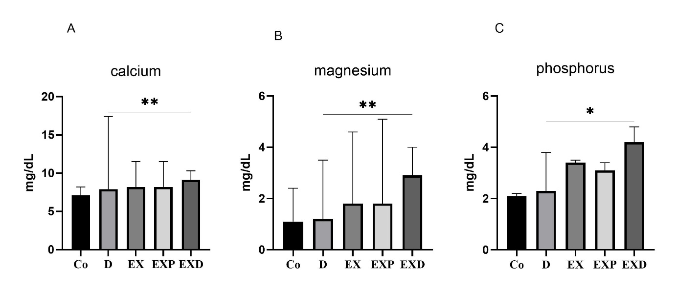
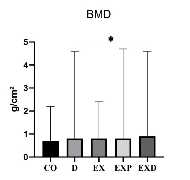
Figure 3. Effects of vitamin D supplementation and Pilates training on bone mineral density (BMD). Data are presented as mean ± SEM (n = 10 per group). Statistical analysis was performed using two-way mixed ANOVA with Bonferroni post-hoc correction. *p ≤ 0.05, **p ≤ 0.01 versus baseline within group; ns, not significant.
Discussion
The findings of the present study demonstrated that eight weeks of Pilates training combined with vitamin D supplementation led to significant improvements in mineral-related biomarkers, increased BDNF levels, and enhanced bone mineral density (BMD) in women with osteosarcoma. The superiority of the combined intervention compared with single interventions suggests potentially beneficial additive effects between the mechanical stimulus of exercise and vitamin D–related biochemical regulation, a concept that has also been highlighted in recent systematic reviews (14, 15).
From a biological perspective, vitamin D contributes to calcium–phosphorus homeostasis and bone remodeling through multiple physiological processes, including calcium absorption and osteoblast-related regulation (16, 17). In vitamin D–deficient populations, correction of deficiency may support a more favorable physiological environment for bone-related adaptation. Given that the majority of participants in this study presented with low baseline vitamin D levels, supplementation may have contributed to improved mineral-related outcomes; however, because post-intervention serum 25(OH) D and mechanistic biomarkers were not directly measured, these interpretations should be considered cautiously.
Exercise may also contribute to bone adaptation through mechanical loading and musculoskeletal stimulation. Although Pilates is generally considered a moderate-intensity modality, its controlled resistance, postural demands, and neuromuscular emphasis may provide sufficient physiological stimulus to support musculoskeletal health, particularly in individuals with reduced physical capacity (18-20). This may help explain why exercise-containing groups demonstrated more favorable outcomes than non-exercise controls.
Some previous studies have reported inconsistent effects of vitamin D supplementation alone on BMD (21, 22), suggesting that supplementation without adequate physical stimulus may have limited structural benefit. In contrast, combined interventions involving exercise and nutritional support may provide broader physiological advantages, particularly in clinically vulnerable populations (14, 15).
The increase in BDNF observed in the combined group represents another potentially relevant finding. Exercise has been associated with elevated circulating BDNF, and vitamin D may influence neurobiological regulation (23, 24). Because BDNF may contribute to neuromuscular function, the observed increase could reflect broader neurophysiological adaptation; however, mechanistic pathways underlying this response were not directly assessed in the present study.
The findings suggest that combining Pilates training with vitamin D supplementation may produce beneficial combined effects on mineral-related outcomes, BDNF, and bone health in women with osteosarcoma. Although vitamin D–related biological pathways, mechanotransduction, and neurotrophic regulation may partially contribute to these outcomes, the present study did not directly assess specific mechanistic pathways such as VDR signaling, Wnt/β-catenin activity, IGF-1, inflammatory markers, oxidative stress, or parathyroid hormone. Therefore, these proposed mechanisms should be interpreted as theoretical hypotheses rather than confirmed physiological explanations.
In patients with osteosarcoma, prior cancer treatment may contribute to impaired bone health, reduced physical function, and rehabilitation challenges (25). Therefore, multidimensional interventions incorporating appropriately supervised exercise and correction of nutritional insufficiencies may represent a potentially valuable supportive rehabilitation strategy. Recent work in exercise oncology similarly emphasizes the importance of integrated supportive interventions for improving physical and skeletal outcomes in cancer survivors (26).
Overall, the present findings support the possibility that combining Pilates training with vitamin D supplementation may improve selected biochemical, neurotrophic, and bone-related outcomes in women with osteosarcoma. However, given the exploratory nature of this study, small sample size, and absence of direct mechanistic measurements, larger and more rigorous trials are required before definitive mechanistic or clinical conclusions can be established.
Summary of Strengths, Limitations, and Future Directions
This study is one of the few to evaluate the combined effects of Pilates training and vitamin D supplementation in women with osteosarcoma using biochemical, neurotrophic, and bone-related measures. The randomized multi-group design and supervised intervention protocol strengthen the comparative value of the findings.
Several limitations should be considered. The short intervention period and modest sample size may limit generalizability and reduce the ability to detect longer-term skeletal adaptations. Inclusion of women only further limits broader applicability. In addition, dietary intake, sunlight exposure, and treatment variability were not fully controlled. The lack of direct pre/post serum 25(OH)D assessment and mechanistic biomarkers also limited biochemical and physiological interpretation.
Future studies should use larger and more diverse samples, longer intervention durations, and longitudinal follow-up. Including direct vitamin D measurements and mechanistic biomarkers would further clarify biological pathways and strengthen clinical relevance.
Conclusion
Combined Pilates training and vitamin D supplementation over eight weeks was associated with improved mineral-related biomarkers, BDNF, and bone mineral density in women with osteosarcoma. These preliminary findings suggest that integrating structured exercise with nutritional support may offer potential rehabilitative value; however, larger and longer-term studies are needed to confirm efficacy and clinical applicability.
Funding
The authors declare no financial support for this research.
Acknowledgments
We thank all participants for their cooperation.
Conflict of Interest
The authors declare no conflicts of interest.
Ethical Considerations
The Research Ethics Committee of Shahid Chamran University of Ahvaz, Iran (Approval No. approved this study IR.SCU.REC.1404.147). Written informed consent was obtained from all participants before enrollment, and all procedures were conducted in accordance with the principles of the Declaration of Helsinki. The ethics approval is publicly available at https://ethics.research.ac.ir/IR.SCU.REC.1404.147.
References
- 1. Ottaviani G, Jaffe N. The epidemiology of osteosarcoma. Pediatric and adolescent osteosarcoma. 2009:3-13. https://doi.org/10.1007/978-1-4419-0284-9_1
- 2. Rosewarne E, Santos JA, Hart AC, Trieu K, Tekle D, Ide N, et al. Review of national nutrition standards with salt-related criteria for publicly funded institutions around the world. Nutrition Reviews. 2023;81(6):647-57. https://doi.org/10.1093/nutrit/nuac080
- 3. Feldman D, Krishnan AV, Swami S, Giovannucci E, Feldman BJ. The role of vitamin D in reducing cancer risk and progression. Nature reviews cancer. 2014;14(5):342-57. https://doi.org/10.1038/nrc3691
- 4. Nasimi N, Jamshidi S, Askari A, Zolfaghari N, Sadeghi E, Nouri M, et al. Effect of vitamin D supplementation or fortification on bone turnover markers in women: a systematic review and meta-analysis. British Journal of Nutrition. 2024;131(9):1473-87. doi 10.1017/S0007114524000060
- 5. Zhao J-G, Zeng X-T, Wang J, Liu L. Association between calcium or vitamin D supplementation and fracture incidence in community-dwelling older adults: a systematic review and meta-analysis. Jama. 2017;318(24):2466-82. doi:10.1001/jama.2017.19344
- 6. Blay R, Flores L, Kupzyk K, Waltman N, Lappe J, Mack L, et al. Twelve-month resistance and impact exercise program or risedronate provides a relative benefit to hip bone structure in postmenopausal women: results from a randomized controlled trial. Osteoporosis international. 2024;35(5):877-91. https://doi.org/10.1007/s00198-023-07008-x
- 7. Wolf J. The law of bone remodeling. Berlin, Heidelberg, and New York: Springer. 1986:63-5.
- 8. Singh B, Toohey K. The effect of exercise for improving bone health in cancer survivors A systematic review and meta-analysis. Journal of science and medicine in sport. 2022;25(1):31-40. https://doi.org/10.1016/j.jsams.2021.08.008
- 9. Dinoff A, Herrmann N, Swardfager W, Liu CS, Sherman C, Chan S, et al. The effect of exercise training on resting concentrations of peripheral brain-derived neurotrophic factor (BDNF): a meta-analysis. PloS one. 2016;11(9):e0163037. https://doi.org/10.1371/journal.pone.0163037
- 10. Graser S, Mentrup B, Schneider D, Klein-Hitpass L, Jakob F, Hofmann C. Overexpression of tissue-nonspecific alkaline phosphatase increases the expression of neurogenic differentiation markers in the human SH-SY5Y neuroblastoma cell line. Bone. 2015;79:150-61. https://doi.org/10.1016/j.bone.2015.05.033
- 11. Lisakovska O, Labudzynskyi D, Khomenko A, Isaev D, Savotchenko A, Kasatkina L, et al. Brain vitamin D3-auto/paracrine system in relation to structural, neurophysiological, and behavioral disturbances associated with glucocorticoid-induced neurotoxicity. Frontiers in Cellular Neuroscience. 2023;17:1133400. https://doi.org/10.3389/fncel.2023.1133400
- 12. Schett G. Effects of inflammatory and anti‐inflammatory cytokines on the bone. European journal of clinical investigation. 2011;41(12):1361-6. https://doi.org/10.1111/j.1365-2362.2011.02545.x
- 13. Bosy-Westphal A, Deurenberg P, Müller MJ. Body composition. Introduction in Human Nutrition. 2020.
- 14. Fischer C, Jakob F, Kohl M, Kast S, Von Stengel S, Kerschan-Schindl K, et al. Additive Effects of Exercise and Vitamin D Supplementation (with and without Calcium) on Bone Mineral Density in Older Adults: A Systematic Review and Meta‐Analysis. Journal of Osteoporosis. 2023;2023(1):5570030. https://doi.org/10.1155/2023/5570030
- 15. Marmol-Perez A, Ubago-Guisado E, Rodriguez-Solana A, Gil-Cosano JJ, Martinez-Vizcaino V, Cavero-Redondo I, et al. Effect of exercise on bone health in children and adolescents with cancer during and after oncological treatment: A systematic review and meta-analysis. Frontiers in Physiology. 2023;14:1088740. https://doi.org/10.3389/fphys.2023.1088740
- 16. Bouillon R, Carmeliet G, Verlinden L, van Etten E, Verstuyf A, Luderer HF, et al. Vitamin D and human health: lessons from vitamin D receptor null mice. Endocrine reviews. 2008;29(6):726-76. https://doi.org/10.1210/er.2008-0004
- 17. Li Y, Zhao P, Jiang B, Liu K, Zhang L, Wang H, et al. Modulation of the vitamin D/vitamin D receptor system in osteoporosis pathogenesis: insights and therapeutic approaches. Journal of Orthopaedic Surgery and Research. 2023;18(1):860. https://doi.org/10.1186/s13018-023-04320-4
- 18. Bloomfield SA, Little K, Nelson M, Yingling V. American College of Sports Medicine position stand: physical activity and bone health. Med Sci Sports Exerc. 2004;195(9131/04):3611. DOI: 10.1249/01.MSS.0000142662.21767.58
- 19. Mullath Ullas A, Boamah J, Hussain A, Myrtziou I, Kanakis I. Impact of Dietary Patterns on Skeletal Health: A Systematic Review and Meta-Analysis of Bone Mineral Density, Fracture, Bone Turnover Markers, and Nutritional Status. Nutrients. 2025;17(24):3845. https://doi.org/10.3390/nu17243845
- 20. Robling AG, Turner CH. Mechanical signaling for bone modeling and remodeling. Critical Reviews™ in Eukaryotic Gene Expression. 2009;19(4). 10.1615/CritRevEukarGeneExpr.v19.i4.50
- 21. Bolland MJ, Grey A, Avenell A. Effects of vitamin D supplementation on musculoskeletal health: a systematic review, meta-analysis, and trial sequential analysis. The lancet Diabetes & endocrinology. 2018;6(11):847-58. https://doi.org/10.1016/S2213-8587(26)00032-X
- 22. Reid IR. Effects of vitamin D supplements on bone density. Journal of Endocrinological Investigation. 2015;38(1):91-4. https://doi.org/10.1007/s40618-014-0127-0
- 23. Battaglia S, Nazzi C, Thayer J. Fear-induced bradycardia in mental disorders: Foundations, current advances, future perspectives. Neuroscience & Biobehavioral Reviews. 2023;149:105163. https://doi.org/10.1016/j.neubiorev.2023.105163
- 24. Sturgeon KM, Kok DE, Kleckner IR, Guertin KA, McNeil J, Parry TL, et al. Updated systematic review of the effects of exercise on understudied health outcomes in cancer survivors. Cancer medicine. 2023;12(24):22278-92. https://doi.org/10.1002/cam4.6753
- 25. Horiuchi K, Nemoto R, Mizuno T, Susa M, Chiba K. Prevalence of low bone mineral density and risk of fractures in osteosarcoma and Ewing’s sarcoma survivors: A scoping review. Journal of Bone Oncology. 2023;38:100464. https://doi.org/10.1016/j.jbo.2022.100464
- 26. Campbell KL, Winters-Stone K, Wiskemann J, May AM, Schwartz AL, Courneya KS, et al. Exercise guidelines for cancer survivors: consensus statement from international multidisciplinary roundtable. Medicine and science in sports and exercise. 2019;51(11):2375. https://doi.org/10.1249/MSS.0000000000002116